Quick Links
Digestive Health for Horses
As horse owners we want our horses to be happy, healthy, and thriving. The digestive tract is fundamental to your horse’s overall health and wellbeing – not only is it where horse food is digested and absorbed but it is also an important part of the immune system.
The equine digestive system is incredibly complex, and any imbalances or disruptions in their gut can lead to a variety of health issues, from colic to gastric ulcers. The horse is reliant on the microbial population living in the gut to break down fibre providing a source of slow-release energy – this process is what enables herbivores to thrive on a fibre-based diet and is why looking after the good bugs is so important for good gut health for horses.
The Equine Digestive System
The horse has evolved to utilise a high fibre diet and, when allowed to graze freely, they will spend in the region of 16-18 hours per day eating – this is known as trickle feeding. Domestication and the greater energy demands of horses in work contributed to a change in diet for the horse, with one of the key differences being an increase in starch intake due to more cereal grains being fed. When fed in excessive quantities and at the expense of feeding fibre, increased starch intakes can result in digestive disturbance and a range of health problems. Increased understanding of the link between starch intake and health issues has started a move back to using high fibre feeds of better quality to meet the needs of the modern day horse but there is still a relatively high incidence of issues like colic and gastric ulcers. To try and avoid such problems it is beneficial to understand the digestive process so we can feed more sympathetically.
The Mouth
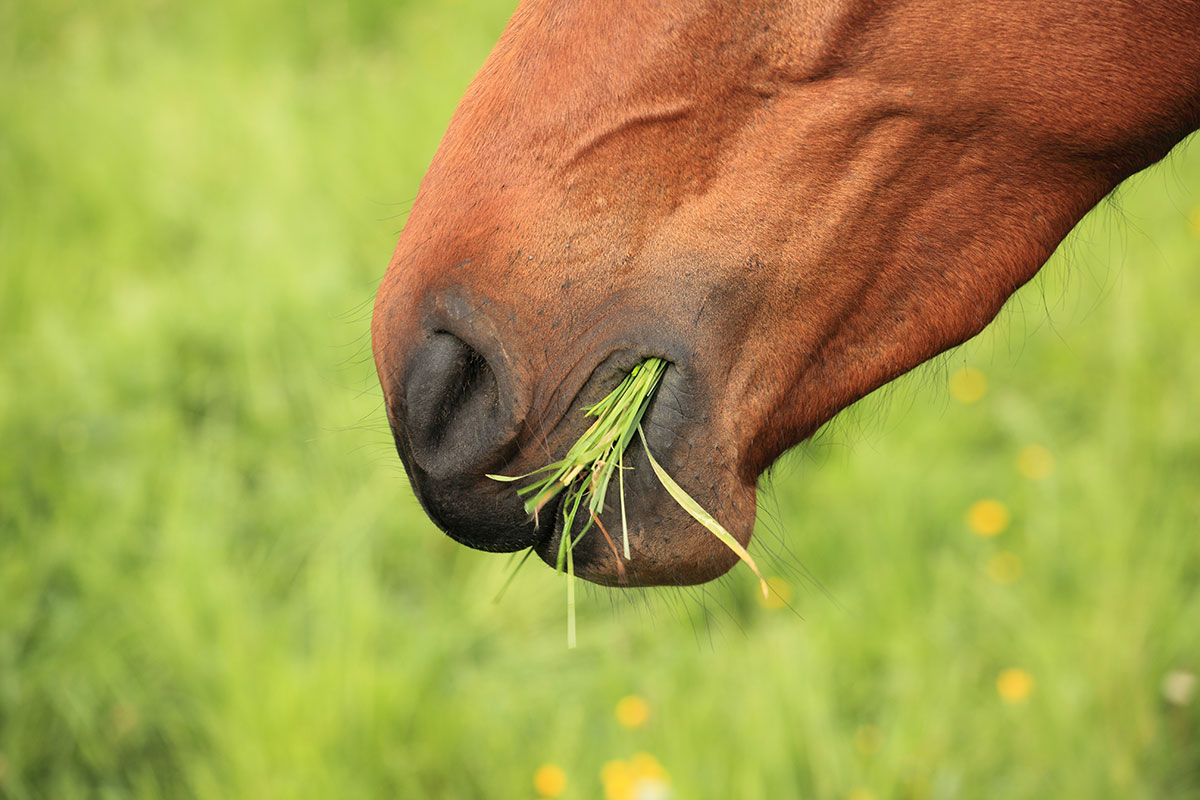
The digestive process starts at the mouth. Horses use their lips to select or avoid different feeds or materials, which is known as prehension. If you watch a horse eating, you will see that their lips are very mobile, which is important for this process. Since horses can’t be sick (the reasons for which are covered later), they need to be very careful about what they are consuming. This becomes very obvious if you have ever tried to hide medication in the feed, as they will often carefully eat everything other than the medicine you want them to consume!
The next step is called mastication, which is the process of chewing. The incisors, the teeth at the front of the mouth, cut or shear the food, before the tongue manoeuvres it to the back of the mouth where it is ground down by the molars into smaller particles. This is known as mechanical digestion. Adult horses have between 36-44 teeth that continue to erupt over the course of their lifetime as the grinding surface is worn away (known as hypsodont teeth). The number of teeth they have is dependent on whether they are male or female, as well as individual variation in whether or not they develop canine and wolf teeth.
The motion that the horse chews will vary when different feeds are eaten, and using high levels of mixes and cubes at the expense of fibre can increase the likelihood of dental irregularities occurring, such as enamel overgrowths or sharp edges (Bonin et al., 2007). In contrast, high fibre diets encourage the horse to use slower, larger movements when chewing, which results in more even wear on the teeth. For more information on dental problems in horses click here.
Consumption times depend on the rate of intake, which includes the time spent chewing. It is not surprising that feeding chopped fibres or forage encourages more chew time compared to cereal based feeds like mixes or cubes. Researchers measured the number of chews taken to eat 1kg of various feed types, and found that it took 3000-3500 chews to eat 1kg of hay, compared to just 832 chews to eat 1kg of oats (Meyer et al., 1975). Ponies need even more chews per kilogram, due to their smaller mouth size. Unsurprisingly, the time taken to consume these quantities also differed greatly, with it taking around four times longer for the horse to eat 1kg of hay compared to the same weight of oats.
The horse has three pairs of salivary glands situated on the sides of the face and adjacent areas of the neck. They differ from humans in that saliva production is not a reflex response. When we smell something nice cooking, we produce saliva automatically. In the horse, however, food has to be present in the mouth and they have to be physically chewing in order to stimulate saliva production. Another difference to humans is that equine saliva contains virtually no digestive enzymes, so it is not until further down the digestive tract that these appear. Saliva still has an important role for the horse though, as it mixes with the feed in the mouth to soften the food and act as a lubricant to aid swallowing. It also contains bicarbonates that buffer acidity within the digestive tract.
It is often quoted that horses produce around 10-12 litres of saliva per day, but in practice this is likely to be highly variable according to how ‘dry’ the feedstuff is and how much chewing is required. The activity of the masseter muscle, which is essentially the horse’s big cheek muscle, has been linked to saliva production and the more active it is, the more saliva is produced. Researchers have shown that eating hay, haylage and chopped fibre is associated with more intense masseter muscle activity in contrast to eating cereals (Vervuert et al., 2013). Using fibre feeds instead of cereals is therefore going to increase the amount of saliva produced, which will increase the acid buffering in the stomach.
The Oesophagus
Once chewed and mixed with saliva, feed is moved to the back of the mouth with the help of the tongue and formed into a bolus where it is swallowed and enters the oesophagus. From this point onwards, the digestive system can be thought of as a long muscular tube with various enlargements along the way. Just as the muscles in your arm or leg contract and relax to move you around, the muscles in the digestive system contract and relax to squeeze the food through the body. This process is known as peristalsis. There are various factors that affect peristalsis; for example, if a horse is frightened or excited, the rate of movement of feed through the digestion system is often increased.
The oesophagus extends between the mouth and the stomach and is approximately 1.2-1.5 metres long. Because the horse has evolved to have their head down grazing, peristalsis in the oesophagus allows the food to go against gravity to move up their neck, and a powerful sphincter muscle at the end of the oesophagus stops food passing back out of the stomach. This means that a horse cannot regurgitate food – if you do see food coming out of a horse’s mouth or nostrils, they are probably choking rather than being sick.
Choke
- An obstruction of the oesophagus.
- The horse can still breathe, but is unable to clear the oesophagus of food as everything is stuck.
- They may appear to be coughing whilst stretching their neck out.
- Although choke may resolve by itself, a vet should be called to aid the clearance of the blockage if necessary.
Preventing choke largely relies on controlling the rate of eating and therefore the particle size swallowed. Horses that are greedy eaters or older horses whose teeth have worn down or fallen out may not fully chew their feed which results in a larger particle size. Less chew time also equals less saliva production and therefore less lubrication to aid swallowing. This is one of the reasons why it is commonly recommended to add chopped fibre when feeding mixes and cubes to slow the rate of intake. Providing easy-to-chew forage replacers may be required for those with poor dentition. More information on feeding forage replacers can be found here.
The Stomach
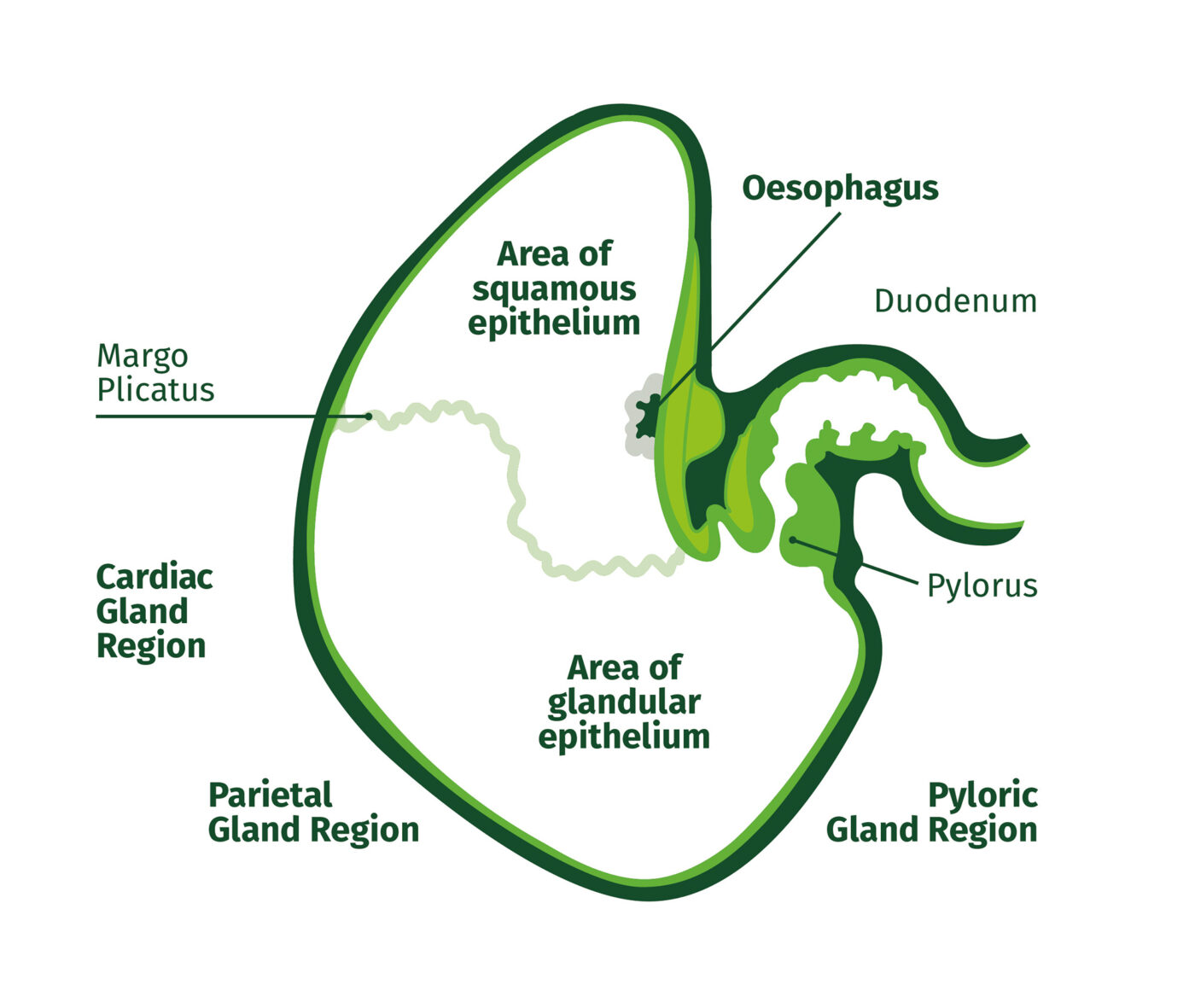
The oesophagus leads to the stomach, which is commonly referred to as being the size of a rugby ball and comprises only around 8-10% of the horse’s total gastrointestinal tract volume. It is one of the enlargements along the digestive system – these enlargements help to slow the rate of passage of food long enough for digestive processes to occur efficiently. The horse is a non-ruminant herbivore, which means that they have a simple stomach similar to a human’s, rather than having four different stomach compartments like a cow.
As the horse has evolved to trickle feed, the stomach has a fairly rigid structure and so cannot stretch to accommodate a large meal. This is in contrast to a carnivore such as a lion who makes one kill, eats as much as possible and then doesn’t eat for a couple of days. The horse is designed to function best on an almost continuous trickle of feed through the digestive system – hence the reason for feeding little and often. The larger the meal, the faster the rate at which the stomach will empty, which does not allow for such efficient digestion. However, highly fibrous feeds like chopped fibres are digested in the hind gut, so we can think of them like hay. For this reason, there’s no need to limit meal size of these feed types, in contrast to cereal based feeds. Food stays in the stomach for around 20 minutes before passing into the small intestine, although it can be longer as the horse’s stomach would never be totally empty when trickle feeding.
The horse’s stomach can be divided into two very distinct regions. Food initially entering the stomach enters the squamous or non-glandular region which is located at the top of the stomach. An area called the margo plicatus marks the change between the squamous to the glandular mucosa at the bottom of the stomach.
Squamous Region
In the top half of the stomach, the horse relies on the presence of fibre to act as a physical barrier to stop gastric acid coming into contact with the squamous mucosa stomach lining. The presence of saliva is also important as it contains bicarbonate which helps to neutralise the acid, and fibre feeds like alfalfa which contain high levels of calcium can further aid in acid buffering. When gastric ulcers occur in the squamous region, this is known as Equine Squamous Gastric Disease (ESGD). High fibre diets are important for reducing the risk of ESGD occurring, in contrast to high starch diets which can increase acidity in the stomach.
Glandular Region
The glandular mucosa is responsible for secreting hydrochloric acid and, as a result, the glandular region typically has a lower pH compared to the squamous region. As horses have evolved to trickle feed, their stomach continually produces acid. The glandular mucosa also secretes mucus and bicarbonate to protect its own lining from the damage that would otherwise occur due to the acid produced. These protection systems may be disrupted which can result in Equine Glandular Gastric Disease (EGGD). The enzymes, pepsin and lipase, are also secreted in this region and these aid in the breakdown of food, with pepsin being responsible for starting to breakdown the structure of proteins in the feed.
More information on ESGD and EGGD can be found here.
The Small Intestine
To exit the stomach, food passes through the pyloric sphincter into the small intestine, which is made up of three regions – the duodenum, jejunum and ileum. It is called the small intestine because it has a small diameter, not because it is short in length – it is actually around 25 metres long which is about the same as two double decker buses.
In contrast, the diameter is only 7-10cm wide – only about the length of a credit card. This is important as it is the site of absorption for many nutrients – if it was a very wide tube then the food would pass through without touching the sides and not much would be absorbed. There are little finger-like projections called villi that line the small intestine to increase the surface area which further help to improve the absorption of nutrients. If the villi were rolled out flat, a horse’s small intestine would be roughly the size of three tennis courts, so this gives us an idea of just how much surface area there is available for nutrient absorption. Research in a range of animals including the horse has shown that high starch diets create a more acidic environment in the digestive tract and this can impact on the size of the villi (Colombino et al., 2022). Smaller villi mean there is less surface area for absorption and less of the immune cells that are also found in gut tissue. Starch levels should be kept as low as possible to optimise gut health and immune function.
It can take anywhere between 30 minutes and four hours for food to pass through the small intestine, and this can be influenced by factors including:
- Feed type – whether it is a mix, cube, chopped fibre or forage
- Meal size – larger meals tend to be pushed through the system quicker
- Feed particle size – how much it has been chewed prior to swallowing
The longer time taken, the more time there is for nutrient absorption to occur.
The nutrients that are broken down and absorbed in the small intestine include protein, fats and oils, most vitamins and minerals, and some soluble carbohydrates like sugar and starch. Bile from the liver is secreted into the small intestine and buffers the pH or acidity of the gut to make conditions more alkaline. This is important to allow digestive enzymes to work and break down the feed, as well as facilitating the movement of the products of digestion across the intestinal wall. The digestive enzymes are located in microvilli which are found at the top of each of the villi throughout the small intestine.
One such digestive enzyme is amylase, which breaks down starch. As horses have evolved to eat fibre, they produce comparatively little amylase compared to other animals and so the digestive system has a limited capacity to utilise starch. Starch is found in high levels in cereals, and excessive quantities of these cause a more rapid transit through the digestive tract which can result in starch reaching the hind gut, where it can cause problems.
Meal size is also very important in affecting how much starch is absorbed in the small intestine. Too large a meal will mean that more starch reaches the hind gut, and so concentrate meal sizes should not weigh more than 400g per 100kg of bodyweight (e.g., 2kg for a 500kg horse), or provide more than 1g of starch per kilogram of bodyweight – whichever is smaller. Starch intake in a 24-hour period should not exceed 2g per kilogram of bodyweight. Dengie’s Starch Calculator can be used to help work this out.
Using fibre as an energy source rather than cereals can be a preferable option because this will support the capacity for nutrient absorption, as the horse’s starch intake is able to remain very low.
Intestinal Parasites
Parasites (worms) can damage the intestinal wall, leaving scar tissue that is less able to absorb nutrients than healthy tissue, meaning that horses will not thrive as well. This may be one reason why some older horses tend to be less efficient at holding weight – they may have years of low grade worm damage which hasn’t necessarily caused them any apparent problems, but the cumulative effect means that after 20 or more years, they are not able to absorb as many nutrients. There is also evidence to suggest that tapeworms in particular are connected to colic incidence (Proudman et al., 1998). Tapeworms attach at the site where the ileum and caecum join, called the ileocaecal junction, and this causes irritation and inflammation of the intestinal lining. In turn, this has an effect on the peristalsis action of the digestive system, increasing the risk of impaction colic, spasmodic colic and intussusception colic, where the small intestine pushes into the caecum. Good pasture management and an appropriate worming programme using faecal egg counts are essential.
The Hind Gut
Also known as the large intestine, the equine hind gut is made up of the caecum, large colon and small colon and is where fibre fermentation takes place.
The caecum is the first part of the hind gut. It is approximately a metre long and is sometimes described as a blind-ended sack, as it is a pouch-like structure that is closed at one end, so feed material has to pass back out very close to where it went in. The caecum has a capacity of 23-35 litres and helps to slow the rate of passage through the digestive system and allow sufficient time for fibre fermentation to occur.
The large colon, given its name by its large diameter of up to 50cm in some areas, is the next part of the hind gut and is around 3-4 metres in length in the average 500kg horse, although it gradually increases in size throughout the horse’s life (Smyth, 1988). It has a capacity of about 80 litres, equivalent to 40 big bottles of soda or 140 pints of milk. This is followed by the small colon, which is approximately three metres long and is the last point at which the horse can absorb anything before faecal balls begin to form and pass through to the rectum.
Mucus lines the colon and offers protection to the colonocytes or gut cells and is also where microbes reside. The colon contains more mucus producing cells than the small intestine as it provides a source of nutrients for bacteria – some of which are very efficient at degrading mucus which is another reason why microbial dysbiosis can be so damaging to health. Studies have shown that when there isn’t enough fibre, bacteria will utilise the mucus as a source of nutrients more and so erode the mucus layer (Desai et al., 2016). This leaves the gut more vulnerable to acidity which compromises the joins between cells; a phenomenon known as leaky gut syndrome. If the gut is leakier or more permeable, it means pathogens and substances that wouldn’t normally escape from the gut into the body can now do so which can cause poor health and disease.
Fibre has a complex structure, and mammals don’t possess the enzymes that are required to break it down. Instead, they rely on millions of bacteria and other micro-organisms like fungi and protozoa in the hind gut to break it down by microbial fermentation. The number of bacterial cells in the horse’s digestive tract is more than 10 times that of all the tissue cells in the whole body, which demonstrates the importance of their role.
Microbial fermentation produces short chain fatty acids (SCFAs), like acetate and butyrate, which are absorbed through the intestine and converted to glucose or fat for energy use or storage, making fibre one of the main energy sources for the horse. Research has shown that diet type can affect the levels of different SCFAs produced in different parts of the gut, with a high fibre diet producing significantly greater levels of butyrate and acetate in the large colon compared to a high starch diet (Raspa et al., 2022). Since butyrate is an important energy source for the cells lining the intestine (which are called colonocytes), a sufficient production of this from fibre fermentation is key for maintaining the function of the intestinal barrier and supporting gut health.
There are several by-products of fermentation including gas and heat:
- Fibre helps to push gas out of the digestive system, and a lack of fibre can often result in gas colic. This is often a problem for older horses that are not able to consume enough fibre due to poor teeth.
- Fibre fermentation also produces heat and this is literally the horse’s central heating system, so feeding plenty of fibre can help to keep the horse warm.
The bacteria that break down the fibre also produce B-vitamins such as biotin. Poor feet can therefore sometimes be an indication that the hind gut is not healthy, as biotin is important for hoof horn integrity. Horses that get very stressed often have loose droppings, as the food passes through the digestive tract too quickly before the water can be absorbed. This increased rate of passage means that bacteria are also carried out of the digestive system, which can mean that B-vitamins are not produced. If a horse has poor feet, is underweight and has a dull coat, providing a digestive aid supplement containing yeast and prebiotics is nearly always beneficial to support fibre digestion alongside a high fibre diet.
Fermenting fibre is not a quick process and feed stays in the large intestine for much longer than it does in any other part of the digestive tract (around 35 hours or longer). How much energy is provided by fibre depends on how much of it can be degraded before it passes into the rectum. Fibre sources such as straw that contain lots of lignin, which gives fibre its structure, are not very digestible and take longer to break down. More digestible fibre sources, including alfalfa and sugar beet, can contribute to a greater extent to the horse’s energy requirements and so are more useful for working horses or those that struggle to maintain weight.
Water is also stored and absorbed from the large intestine, and different feeds and fibres have differing water holding capacities. This is something that endurance riders are very familiar with and is one of the reasons they favour the use of sugar beet. Not only is sugar beet fed soaked and therefore introduces water in to the digestive tract, but as sugar beet pulp is also a very digestible source of fibre, it breaks down easily, releasing the water to aid hydration.
What happens if there is starch overspill from the small intestine?
- Large amounts of sugar and starch that have escaped digestion in the small intestine are rapidly fermented in the hind gut.
- This can lead to changes in the composition of the microbial population, especially if insufficient fibre is fed.
- This results in the death of bacteria that cannot tolerate the more acidic conditions.
- Endotoxins are released along with other factors that cause damage to the lining of the large intestine, effectively making it more “leaky”.
- Toxins and other substances can then end up being absorbed into the blood stream.
- This can trigger problems such as colic and diarrhoea.
As well as keeping starch intake and meal sizes within suitable limits (as discussed above), avoiding any sudden changes to the feeding regime can also help to reduce the likelihood of digestive disturbance (dysbiosis). Fibre-digesting bacteria are important for keeping harmful species of bacteria at bay and thrive in a consistent environment. Sudden changes to the diet can cause bacteria to die off as their environment becomes less hospitable. This can result in the production of toxins which may result in problems such as colic. These changes not only include those to the bucket feed, but changes in grazing management as well, with any adjustments being made gradually over 7-14 days.
Due to the large size and surface area of the equine hind gut, diseases that affect this area of the intestine can result in serious and life threatening complications. For this reason, any changes in normal gut function must be taken seriously.
The Rectum
The last part of the digestive system is the rectum. This is about 30cm long in the horse and acts as the storage area for waste material that hasn’t been absorbed before it is excreted. Horse’s faeces can be a good indicator of digestive health: the number, frequency and consistency should be monitored as they provide a good indication of the horse’s health. If the horse is fed a very coarse, fibrous hay then it is likely that a large proportion will remain undigested. This isn’t a problem for them as they need lots of fibre to maintain gut function – it just means there might be more mucking out to do!
Ancillary Organs
- Liver – produces bile which drains continuously into the small intestine, as horse doesn’t have a gall bladder. Bile contains bile salts which help to break down fats and oils.
- Pancreas – contains lots of digestive enzymes and helps to neutralise the food coming from the stomach to aid enzyme activity. The pancreas also produces hormones such as glucagon which is produced in response to a drop in blood glucose and stimulates the breakdown of glycogen. A rise in blood glucose results in the release of insulin which stimulates the storage of glucose.
- Kidney – controls the water potential of the blood and so high concentrations of certain substances will be excreted through the urine. For example, nitrogenous waste from the breakdown of amino acids will be excreted through the urine.
Top Tips for Gut Health
- Feed little and often
- Feed plenty of fibre
- Avoid making sudden changes to the diet
- Keep up with regular dental checks and ensure that those with poor dentition have access to a fibre type they can chew easily
- Minimise stress where possible
- Avoid using excessive levels of cereal based feeds
- Use high-quality feed ingredients
- Ensure the horse has access to water
- Use feeds containing alfalfa to aid acid buffering in the stomach
- Monitor changes in the faeces as this will help to identify if there is a problem
- Implement a suitable parasite control programme
References
- Bonin, S.J., Clayton, H.M., Lanovaz, J.L. and Johnston, T. (2007) Comparison of mandibular motion in horses chewing hay and pellets. Equine Veterinary Journal, 39(3), pp.258-262.
- Colombino, E., Raspa, F., Perotti, M., Bergero, D., Vervuert, I., Valle, E. and Capucchio, M.T. (2022) Gut health of horses: effects of high fibre vs high starch diet on histological and morphometrical parameters. BMC Veterinary Research, 18(1), p.338.
- Desai, M.S., Seekatz, A.M., Koropatkin, N.M., Kamada, N., Hickey, C.A., Wolter, M., Pudlo, N.A., Kitamoto, S., Terrapon, N., Muller, A. and Young, V.B. (2016). A dietary fiber-deprived gut microbiota degrades the colonic mucus barrier and enhances pathogen susceptibility. Cell, 167(5), pp.1339-1353.
- Meyer, H., Ahiswedeu, L. and Reinhardt, H.J. (1975) Studies on the duration of feeding, masticatory frequency and mincing of feed in horses. DTW. Deutsche Tierarztliche Wochenschrift, 82(2), pp.54-58.
- Proudman, C.J., French, N.P. and Trees, A.J. (1998) Tapeworm infection is a significant risk factor for spasmodic colic and ileal impaction colic in the horse. Equine Veterinary Journal, 30(3), pp.194-199.
- Raspa, F., Vervuert, I., Capucchio, M.T., Colombino, E., Bergero, D., Forte, C., Greppi, M., Cavallarin, L., Giribaldi, M., Antoniazzi, S. and Cavallini, D. (2022) A high-starch vs. high-fibre diet: Effects on the gut environment of the different intestinal compartments of the horse digestive tract. BMC Veterinary Research, 18(1), pp.1-11.
- Smyth, G.B. (1988) Effects of age, sex, and post mortem interval on intestinal lengths of horses during development. Equine Veterinary Journal, 20(2), pp.104-108.
- Vervuert, I., Brüssow, N., Bochnia, M., Cuddeford, D. and Coenen, M. (2013) Electromyographic evaluation of masseter muscle activity in horses fed (i) different types of roughage and (ii) maize after different hay allocations. Journal of Animal Physiology and Animal Nutrition, 97(3), pp.515-521.